
Retrolisthesis: Symptoms, Causes and Treatment
Retrolisthesis is a spinal condition in which one vertebra shifts backward relative to the vertebra directly beneath it. The term can sound alarming when it appears unexpectedly on an X-ray or MRI report, but the imaging finding does not automatically mean that severe spinal damage or surgery is inevitable. Some people with lumbar retrolisthesis experience lower back pain, stiffness, sciatica, or reduced mobility, while others have little or no noticeable discomfort. Symptoms often depend on other changes occurring around the affected spinal segment, including disc degeneration, facet joint arthritis, spinal stenosis, or nerve compression. Understanding the entire clinical picture is therefore more useful than focusing only on the amount of backward vertebral slippage.
Retrolisthesis is less extensively studied than the more familiar forward vertebral slippage called anterolisthesis or spondylolisthesis. Recent research continues to show that retrolisthesis commonly occurs alongside degenerative changes and altered spinal alignment rather than functioning as an isolated disease in every patient. A 2024 cross-sectional study of people attending spine clinics found retrolisthesis in 17.1% of participants, with L3-L4 being the most frequently affected level, although this percentage should not be interpreted as prevalence in the general population. Most cases are treated according to symptoms, neurological findings, spinal stability, and accompanying conditions. This guide explains retrolisthesis symptoms, causes, diagnosis, exercises, treatment options, prognosis, and warning signs that deserve prompt medical attention.
What Is Retrolisthesis?
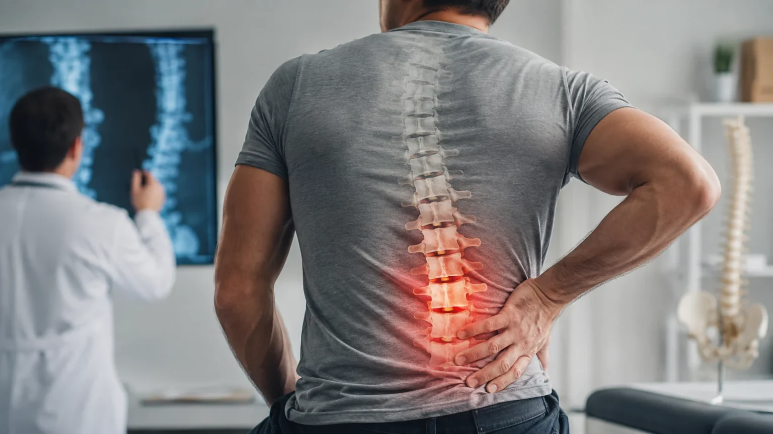
Retrolisthesis describes posterior translation of one vertebral body relative to the adjacent vertebra underneath it. In simple terms, one of the bones forming the spine sits slightly farther backward than expected when compared with the neighboring bone. It is considered a form of vertebral displacement and is essentially the opposite directional movement of anterolisthesis, where a vertebra moves forward. Retrolisthesis is most commonly discussed in the lumbar spine because degenerative lower-back problems frequently lead to imaging, although backward displacement can theoretically occur at other spinal levels. The finding may be discovered on a standing X-ray, CT scan, or MRI performed because of back pain, leg symptoms, or another reason entirely.
The vertebrae are normally held in alignment by intervertebral discs, facet joints, ligaments, muscles, and other supporting tissues. These structures work together to permit controlled movement while preventing excessive translation between neighboring spinal bones. As discs lose height or facet joints undergo degenerative changes, the mechanics of a spinal segment can gradually change. A vertebra may then shift slightly backward as part of that altered mechanical relationship. Research examining degenerative lumbar disease suggests that retrolisthesis may sometimes act as a compensatory mechanism that moves the body’s gravity axis posteriorly when sagittal spinal balance has changed. This means retrolisthesis can sometimes reflect the spine’s broader alignment rather than a single isolated injury.
Retrolisthesis should not automatically be considered equivalent to spinal instability. An imaging report may describe a small backward displacement even when the vertebral segment remains relatively stable during movement and the patient has few symptoms. Conversely, a modest-looking slip may coexist with disc herniation or narrowing around a nerve root that produces significant leg pain. This is why doctors generally evaluate the patient’s symptoms, physical examination, neurological findings, and other imaging abnormalities rather than judging severity from one measurement alone. A study involving patients with L5-S1 disc herniation found that retrolisthesis itself was not significantly associated with worse baseline pain or function in that particular group.
Retrolisthesis can also appear alongside degenerative disc disease, facet joint degeneration, scoliosis, spinal stenosis, or another type of vertebral slippage. Researchers studying patients with lumbar spondylolytic spondylolisthesis have documented degenerative changes involving both discs and facet joints at adjacent levels where retrolisthesis was present. These overlapping abnormalities matter because a patient’s pain may arise from the disc, joints, compressed nerves, muscles, or several structures simultaneously. Consequently, treating the imaging finding alone without determining the likely pain generator may lead to unrealistic expectations. Modern management therefore focuses more heavily on function and symptoms than on attempting to make every spinal image appear anatomically perfect.
Another reassuring point is that finding retrolisthesis does not automatically mean that the spine will continue slipping backward. Degenerative spinal conditions may remain stable for long periods, and the natural history varies widely between individuals. Many people can manage symptoms with physical therapy, exercise, activity modification, and appropriate pain treatment rather than surgery. Follow-up becomes more important when symptoms progressively worsen, neurological changes develop, or imaging suggests meaningful instability or nerve compression. Anyone reading the word “retrolisthesis” on an imaging report should therefore discuss what the finding means in their specific case. The clinical importance depends on where it occurs, how much translation is present, what surrounding structures look like, and whether those changes actually correspond with symptoms.
Retrolisthesis Symptoms
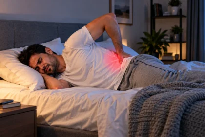
Lower back pain is one of the most common complaints associated with symptomatic lumbar retrolisthesis, although the vertebral displacement may not always be the direct source of pain. The discomfort can feel dull, aching, stiff, or occasionally sharp depending on activity and the structures involved. Some people notice symptoms after prolonged sitting, standing, lifting, bending, or other activities that place repeated stress on the lumbar spine. Pain may remain concentrated around the affected area or spread into the buttocks and upper legs. Because similar symptoms occur with muscle strain, degenerative disc disease, arthritis, and many other back conditions, pain alone cannot confirm retrolisthesis. Imaging and clinical evaluation are needed when identifying the underlying structural changes would affect management.
Back stiffness and reduced mobility can accompany pain, particularly when degenerative changes involve the intervertebral discs or facet joints. A person may notice difficulty bending forward, rotating the trunk, standing upright after sitting, or completing activities that previously felt comfortable. Protective muscle tightening can make the lower back feel rigid even when the actual vertebral translation is relatively small. People sometimes describe morning stiffness or discomfort after remaining in one position for an extended period. Gradual strengthening and movement can help many mechanical back conditions, but an exercise program should be adapted when symptoms are severe or neurological problems are present. The presence of stiffness does not necessarily indicate that the vertebra is actively continuing to slip.
Nerve-related symptoms may develop when degenerative changes, disc problems, or altered spinal alignment narrow the space available for a spinal nerve. Sciatica can produce pain traveling from the lower back or buttock down the thigh and sometimes into the calf or foot. The discomfort may feel burning, electric, shooting, or shock-like rather than simply aching, and numbness or tingling can occur along the same pathway. Muscle weakness can also develop when nerve compression becomes more significant. These symptoms are not unique to retrolisthesis and frequently occur with herniated discs or spinal stenosis. Mayo Clinic notes that sciatica can include leg pain, tingling, numbness, and weakness, usually involving one side of the body.
Walking or standing tolerance may also decrease when retrolisthesis occurs with spinal stenosis or other degenerative narrowing. Some people develop heaviness, aching, tingling, or weakness in the legs after standing or walking for a certain distance, a pattern sometimes associated with neurogenic claudication. Others simply notice that the back fatigues quickly and that they need to change positions more frequently. Symptoms may improve when sitting or leaning slightly forward if spinal stenosis is contributing, although individual patterns vary. Difficulty walking deserves medical assessment when it is new, progressively worsening, or associated with weakness or balance problems. Determining whether symptoms come from nerves, joints, discs, muscles, or another condition is essential before deciding on treatment.
Many people with vertebral slippage have few symptoms or discover the abnormality incidentally during imaging for another problem. This distinction is important because radiological abnormalities become more common with age and do not always correspond closely with how much pain someone feels. Research into retrolisthesis remains more limited than research into forward spondylolisthesis, and existing studies do not support assuming that every backward slip causes major disability. A clinician should therefore ask whether the location and associated abnormalities plausibly explain the patient’s symptoms. When retrolisthesis is an incidental finding without pain, neurological deficits, or instability, treatment aimed specifically at the slip may not be necessary.
What Causes Retrolisthesis?
Age-related spinal degeneration is one of the most important settings in which retrolisthesis develops. Intervertebral discs normally act as cushions and help maintain the spacing and alignment between neighboring vertebrae. Over time, discs can lose hydration, elasticity, and height, while nearby facet joints may develop arthritic changes. These changes alter how forces are transferred through the spinal motion segment and can allow small amounts of abnormal translation. Retrolisthesis has therefore frequently been described in patients with degenerative lumbar spinal disorders. Aging itself does not guarantee that someone will develop painful retrolisthesis, but degenerative changes become more common over time and can create the mechanical environment in which backward vertebral displacement appears.
Changes in spinal balance may also contribute to retrolisthesis. The human spine constantly adjusts to keep the head and trunk balanced over the pelvis while a person stands and walks. If lumbar curvature or pelvic alignment changes, certain vertebral levels may shift as part of a compensatory response. A study of degenerative lumbar spine patients found associations between retrolisthesis and sagittal alignment characteristics, with the authors proposing that backward translation may sometimes help move the gravity axis posteriorly. This concept explains why retrolisthesis should not always be viewed simply as a bone that has randomly “slipped out of place.” In some people, it may represent one component of a more complex change in spinal mechanics.
Disc degeneration and facet joint changes can work together rather than appearing independently. The intervertebral disc carries part of the spinal load, while paired facet joints behind the vertebral bodies guide movement and contribute to stability. When disc height decreases, the load and movement pattern across the facet joints can change. Researchers examining spinal segments adjacent to spondylolytic spondylolisthesis found relationships between retrolisthesis and degenerative changes at the disc and facet joints. This interconnected process is one reason symptoms attributed to retrolisthesis may actually arise from multiple structures. Treatment often targets mobility, strength, inflammation, nerve irritation, and functional limitations instead of trying to address the backward displacement in isolation.
Trauma can occasionally contribute to vertebral displacement, particularly when substantial forces damage discs, ligaments, joints, or bones that normally stabilize the spine. However, a sudden retrolisthesis after a significant fall, collision, or other injury should be evaluated differently from gradual degenerative retrolisthesis. Severe trauma raises concern for fractures or ligament injuries that may require urgent imaging and specialist assessment. Mild back pain after ordinary activity should not automatically be interpreted as evidence that a vertebra suddenly shifted backward. The history of how symptoms started is therefore clinically useful. Sudden severe pain following major trauma, especially when accompanied by weakness, numbness, or difficulty walking, warrants prompt medical evaluation rather than relying solely on self-treatment.
Retrolisthesis risk cannot usually be reduced to one lifestyle factor, and people should be cautious about claims that poor posture alone “causes” a vertebra to slip. Prolonged inactivity, reduced muscle conditioning, repetitive loading, occupational strain, excess body weight, smoking, and poor movement habits may contribute to broader spinal health problems, but their relationship with a specific retrolisthesis is not always straightforward. Current research remains comparatively limited, and associations found in spine-clinic populations do not prove that a particular behavior directly caused the displacement. A newer radiographic study found retrolisthesis most frequently at L3-L4 and associated it with certain spinal alignment parameters. The practical goal is therefore improving overall spinal function and reducing modifiable contributors to persistent back symptoms.
How Retrolisthesis Is Diagnosed
Diagnosis usually begins with a detailed history rather than imaging alone. A clinician may ask where pain occurs, whether it travels into the legs, what movements worsen or relieve symptoms, and whether numbness, tingling, weakness, or walking problems are present. The timing of symptoms can also provide clues, particularly when pain followed trauma or developed gradually over several years. Questions about previous spinal problems, osteoporosis risk, cancer history, infection risk, medications, and other medical conditions help identify alternative causes of back pain. This step is important because a person can have retrolisthesis on an image while their actual pain comes from a different structure or condition. Diagnosis therefore involves correlating the radiological finding with the patient’s clinical presentation.
A physical examination may assess posture, spinal range of motion, areas of tenderness, gait, muscle strength, reflexes, and sensation. When nerve-root irritation is suspected, clinicians may perform tests designed to reproduce radiating leg symptoms or identify neurological deficits. Hip movement may also be checked because hip disorders can produce pain that resembles lumbar spine problems. The examination can help determine whether symptoms are primarily mechanical, neurological, or potentially caused by another medical condition. Progressive weakness or major sensory changes usually raise greater concern than uncomplicated localized lower-back discomfort. No single physical examination maneuver confirms retrolisthesis, however, because identifying actual vertebral translation requires imaging.
Plain X-rays can show the alignment of vertebral bodies and make a backward translation visible, particularly on a lateral view of the spine. Standing radiographs may provide useful information because spinal alignment under normal weight-bearing conditions can differ from alignment while lying down. In selected cases, flexion and extension X-rays may be used to examine how one vertebra moves relative to another and whether meaningful dynamic instability exists. Measurements can describe the amount of displacement, but numbers should be interpreted alongside symptoms and other structural abnormalities. Not every person with uncomplicated low back pain requires an X-ray. NICE and the American College of Radiology both advise against routine imaging for uncomplicated low back pain without red flags because imaging often does not change initial management.
MRI becomes particularly useful when nerve compression, disc disease, spinal stenosis, or another soft-tissue abnormality is suspected. Unlike a standard X-ray, MRI can show intervertebral discs, nerve roots, the spinal canal, ligaments, and other soft tissues surrounding the vertebrae. A clinician may order MRI when symptoms persist despite appropriate conservative care, when significant neurological findings are present, or when the result would influence treatment decisions. MRI can also help determine whether the symptoms attributed to retrolisthesis are better explained by a herniated disc or foraminal narrowing. Imaging findings are still not interpreted in isolation because many degenerative changes appear in people without pain. The most useful MRI is one that answers a clinically relevant question.
CT scanning may be helpful when detailed evaluation of bone anatomy is needed, particularly after trauma, when fracture is suspected, or when MRI cannot provide the necessary information. Additional tests may be ordered if clinicians suspect infection, inflammatory disease, osteoporosis, malignancy, or another systemic cause of spinal symptoms. The diagnostic process is therefore broader than simply proving that retrolisthesis exists. Once the backward displacement has been identified, the more important questions are whether it is stable, whether nerves are affected, and whether another abnormality better explains the symptoms. A clear diagnosis helps prevent unnecessary treatment of incidental imaging findings. It also allows conservative therapy or specialist referral to be targeted toward the actual source of pain and functional limitation.
Retrolisthesis Treatment Without Surgery
Most people with mild or moderate symptoms are initially treated conservatively rather than undergoing surgery solely because retrolisthesis appears on an imaging report. The treatment plan depends on whether symptoms are primarily localized back pain, nerve-related leg pain, stiffness, or a combination of these problems. Remaining reasonably active is generally preferred to prolonged bed rest for uncomplicated mechanical low back pain. NICE recommends self-management advice and continuing normal activities when possible, with exercise programs considered according to the person’s needs and abilities. Short periods of modifying particularly aggravating tasks may still be useful during an acute flare. The long-term objective is typically to restore movement, strength, confidence, and function without repeatedly provoking symptoms.
Physical therapy is frequently an important part of conservative retrolisthesis treatment. A physical therapist can assess spinal movement, hip mobility, core control, leg strength, posture, and activities that repeatedly trigger pain. Treatment may include progressive strengthening, mobility exercises, aerobic activity, and instruction on safer lifting or movement strategies. Programs should be individualized because an exercise that feels comfortable for one patient may aggravate sciatica or stenosis symptoms in another. NICE supports exercise for low back pain and sciatica and considers manual therapy appropriate only when incorporated into a broader treatment package that includes exercise. Passive treatments alone are unlikely to address all of the functional limitations that accompany persistent mechanical back pain.
Medication may provide temporary symptom relief while a person remains active and participates in rehabilitation. Nonsteroidal anti-inflammatory drugs, or NSAIDs, may be considered for low back pain when medically appropriate, but they carry gastrointestinal, kidney, cardiovascular, and other risks. NICE recommends considering individual risk factors and using the lowest effective dose for the shortest appropriate period when NSAIDs are prescribed. People with kidney disease, gastrointestinal ulcers, cardiovascular problems, certain medications, or other health concerns should discuss pain medicines with a clinician or pharmacist rather than assuming an over-the-counter product is safe. Medication can reduce discomfort, but it does not physically return a displaced vertebra to a different position.
Injections are sometimes discussed when retrolisthesis occurs alongside severe nerve-related pain, but their role depends on the actual source of symptoms. Routine spinal injections are not recommended simply for nonspecific low back pain. NICE does, however, state that epidural local anesthetic and steroid injections may be considered for acute and severe sciatica in selected patients. Such procedures are intended to address inflammation and nerve pain rather than permanently correct vertebral alignment. The benefits may also be temporary, meaning injections should usually form part of a broader management strategy. A specialist should determine whether imaging findings and symptoms make a targeted procedure reasonable.
Lifestyle adjustments can support treatment without turning everyday movement into something to fear. Breaking up long periods of sitting, gradually increasing walking or other aerobic activity, improving sleep habits, and using better lifting technique may reduce repeated aggravation. Weight management can be helpful for some people because higher loads may increase stress on already symptomatic spinal structures, although weight alone should never be presented as the sole cause of pain. Smoking cessation is also relevant to broader disc and bone health. The goal is not to achieve “perfect posture” every minute but to build tolerance for normal movement and avoid repeatedly stressing the back beyond its current capacity. Persistent symptoms should lead to reassessment rather than endless restriction of activity.
Exercises and Daily Management for Retrolisthesis
Exercise for retrolisthesis should generally focus on function rather than attempting to force a vertebra physically back into position. Core-strengthening exercises can improve control of the trunk and help muscles share loads that otherwise fall heavily on painful spinal structures. Depending on the individual, a therapist may use exercises such as abdominal bracing, modified bridges, bird-dog variations, controlled dead-bug movements, or other stabilization activities. The specific exercise matters less than whether it can be performed with good control and progressively increased without causing significant symptom worsening. Pain that radiates farther down the leg, new numbness, or weakness during exercise deserves reassessment. A personalized program is particularly important when spinal stenosis, disc herniation, osteoporosis, or previous surgery is also present.
Hip and leg strength can be just as important as abdominal strength when managing persistent lower-back symptoms. Strong gluteal and leg muscles allow everyday tasks such as walking, climbing stairs, lifting, and rising from a chair to be distributed across more than the lumbar spine. Limited hip movement may cause a person to compensate by repeatedly bending or rotating through the lower back. A rehabilitation plan can therefore include hip strengthening, hamstring or hip-flexor mobility work, squatting progressions, and balance exercises when appropriate. Stretching should feel controlled rather than aggressive. Someone does not need extreme spinal flexibility to manage retrolisthesis successfully, and forceful stretching into painful positions can sometimes aggravate irritated joints or nerves.
Walking is often a practical way to reintroduce regular activity because intensity and duration are easy to adjust. Someone with significant symptoms might initially tolerate several short walks better than one long session and can gradually increase duration as pain and endurance improve. Cycling, swimming, water exercise, or other low-impact cardiovascular activities may also be reasonable alternatives depending on personal comfort. NICE encourages continuation of normal activities and the use of appropriate exercise programs for low back pain with or without sciatica. There is rarely one universally “best” exercise for every case of retrolisthesis. Consistency with a tolerable program is generally more useful than repeatedly changing exercises in search of a single corrective movement.
Daily movement habits can reduce unnecessary irritation without requiring rigid posture rules. When lifting, keeping the object reasonably close to the body, using the hips and knees, and avoiding sudden uncontrolled twisting under heavy load can make the task easier on the back. People who sit for long periods may benefit from changing position or standing periodically rather than trying to maintain one supposedly perfect seated position for hours. Workstations can be adjusted so frequently used objects are comfortably reachable and the person can vary posture throughout the day. Gradual exposure is also important because avoiding all bending and lifting can reduce physical capacity over time. Rehabilitation should help people return safely to meaningful activity rather than convince them that their spine is fragile.
Activities that clearly provoke severe symptoms may need temporary modification, particularly during an acute flare, but permanent restrictions are not automatically required. Heavy lifting, repeated high-impact loading, or extreme ranges of spinal motion may be reintroduced gradually when strength, confidence, and symptoms permit. People involved in demanding work or sport may benefit from professional guidance on technique and progressive loading. Braces and corsets should not become routine substitutes for active rehabilitation in uncomplicated chronic low back pain; NICE specifically recommends against routinely offering belts or corsets for low back pain management. The most sustainable approach combines symptom awareness with progressive strengthening and normal movement instead of long-term dependence on passive support.
When Is Surgery Needed for Retrolisthesis?
Surgery is not automatically required because an imaging report mentions retrolisthesis. Most surgical decisions are based on the condition producing symptoms, the degree of neurological compromise or instability, and whether appropriate nonsurgical care has failed. A patient whose main problem is a compressed nerve may require a different procedure from someone with substantial mechanical instability. Surgeons therefore review X-rays or MRI findings alongside symptoms, neurological examination results, and functional limitations before recommending an operation. The presence of several degenerative abnormalities can make this decision more complex because the painful structure must be identified as accurately as possible. Treating an incidental backward slip surgically would not necessarily improve pain originating from an unrelated source.
Persistent sciatica caused by clearly demonstrated nerve compression is one situation in which surgical evaluation may become appropriate. NICE recommends considering spinal decompression when nonsurgical treatment has not improved pain or function and radiological findings are consistent with the patient’s sciatic symptoms. Decompression involves creating more space for affected nerves by removing or reshaping structures responsible for compression. Whether decompression alone is adequate depends on spinal stability and the specific pathology. Some patients can obtain nerve relief without fusion, while others may require stabilization when removing compressive structures would create or worsen instability. A spine surgeon should explain why the proposed operation matches the individual’s symptoms and imaging.
Spinal fusion may be considered in selected patients when clinically meaningful instability accompanies severe symptoms or when stabilization is necessary as part of treatment for another spinal disorder. Fusion connects two or more vertebrae so that motion at the problematic segment is reduced while bone gradually grows across the treated area. Screws, rods, cages, or bone grafts may be used depending on the surgical approach. Fusion is a major operation with potential risks including infection, bleeding, nerve injury, nonunion, and degeneration at adjacent levels. It should therefore not be viewed as a routine treatment for every retrolisthesis. The expected benefit must outweigh those risks and should be based on a clearly defined structural problem.
Surgical decisions also consider how strongly symptoms interfere with everyday life. Someone with persistent severe leg pain, progressive weakness, limited walking ability, or inability to perform normal activities despite well-conducted conservative treatment may have a stronger reason for specialist evaluation than someone with occasional manageable back stiffness. Duration of symptoms matters, but there is no universal waiting period appropriate for every patient. Severe or progressive neurological problems can justify faster intervention, whereas stable mechanical pain may be managed conservatively for considerably longer. Surgical consultation does not obligate a person to undergo an operation. It provides an opportunity to clarify the diagnosis, expected natural history, available alternatives, potential benefits, and realistic recovery expectations.
Emergency surgery is different from elective surgery because neurological function may be at immediate risk. Compression of the cauda equina, a bundle of nerves at the bottom of the spinal canal, can cause new bladder or bowel dysfunction, numbness around the genitals or inner thighs, and significant leg weakness. Mayo Clinic identifies bladder or bowel problems and saddle anesthesia as emergency warning signs because delayed treatment can result in lasting neurological damage. Retrolisthesis itself does not mean cauda equina syndrome is present, and the complication is uncommon. However, anyone with severe back or leg symptoms plus these neurological changes should seek emergency medical assessment instead of waiting for a routine appointment.
Is Retrolisthesis Serious?
For many people, retrolisthesis is not a dangerous condition and may represent a relatively minor component of age-related spinal degeneration. The word can appear dramatic on an imaging report, but the seriousness of the condition depends much more on symptoms and associated abnormalities than on the label itself. A small stable displacement without significant pain or neurological findings may require little more than observation and general back care. Even symptomatic cases are often approached first with exercise-based rehabilitation and other conservative measures. Studies of lumbar retrolisthesis also highlight that its relationship with pain and disability is not always straightforward. This makes individualized interpretation essential.
Retrolisthesis becomes more clinically significant when it is associated with substantial spinal stenosis, disc herniation, instability, or nerve compression. In these situations, the main concern may actually be the effect on surrounding nerves rather than the backward displacement itself. Persistent numbness, progressive muscle weakness, deteriorating walking ability, or severe radiating leg pain deserve medical assessment. A clinician can determine whether symptoms correlate with the level where retrolisthesis appears or whether another spinal abnormality provides a better explanation. Imaging can look severe while symptoms remain modest, and the reverse can also happen. Treatment should therefore focus on the person rather than trying to make treatment intensity match the appearance of the scan.
The degree of retrolisthesis also does not necessarily predict exactly how someone will feel in the future. Some spinal degenerative changes progress, whereas others remain relatively stable over many years. The newer radiographic literature continues to examine relationships between retrolisthesis, age, spinal geometry, and sagittal alignment, but there is still less evidence about retrolisthesis than about more common forms of degenerative spondylolisthesis. This uncertainty makes strong claims about guaranteed progression inappropriate. Monitoring is usually guided by changes in symptoms rather than repeated imaging on an arbitrary schedule. If function is improving and neurological findings remain stable, frequent scans may provide little additional benefit.
People can improve significantly even when follow-up imaging would still show some degree of vertebral translation. Conservative treatment does not necessarily “reverse” retrolisthesis anatomically, but it can improve muscle capacity, mobility, nerve tolerance, pain, and everyday function. This distinction matters because patients sometimes assume successful treatment must result in the vertebra returning to an ideal position. For many degenerative musculoskeletal conditions, clinical improvement is a more realistic treatment goal than radiological perfection. A strong and well-conditioned spine can function well despite structural changes visible on imaging. Progress is therefore better measured by activities such as walking, working, sleeping, lifting, exercising, and participating in daily life with fewer limitations.
The prognosis becomes more concerning when neurological symptoms progress or severe pain remains uncontrolled despite appropriate care. New weakness, major sensory loss, bladder or bowel dysfunction, fever with severe back pain, unexplained weight loss, or pain after significant trauma requires a different level of attention than ordinary chronic stiffness. Serious underlying disease is uncommon compared with routine mechanical low back pain, but identifying warning signs is important. NICE advises clinicians to consider alternative pathology such as cancer, infection, trauma, or inflammatory disease when the history suggests it. Retrolisthesis seen on an X-ray should never prevent investigation of another condition when symptoms do not fit the expected mechanical pattern.
When to See a Doctor for Retrolisthesis
A routine medical appointment is reasonable when lower-back pain lasts for several weeks, repeatedly returns, or significantly limits work, sleep, exercise, or everyday activities. An evaluation is also worthwhile if pain regularly travels into the buttock or leg or if numbness and tingling begin appearing. These symptoms may indicate nerve irritation from a disc problem, spinal stenosis, or another condition occurring alongside retrolisthesis. The clinician can determine whether further imaging, physical therapy, medication, or referral is appropriate. Mild uncomplicated back pain frequently improves without extensive testing, so seeking professional advice does not automatically mean that an MRI or surgical consultation will be required.
Progressive muscle weakness deserves more urgent evaluation than stable mild pain. A person might notice difficulty lifting the front of the foot, repeatedly tripping, trouble standing on the toes, or a leg that feels less reliable than before. New weakness can indicate clinically important nerve compression and should not simply be treated with additional stretching or massage. Sudden neurological symptoms after a major injury require particularly prompt assessment. Mayo Clinic advises immediate care for sudden numbness or muscle weakness associated with severe sciatic symptoms. Early evaluation allows clinicians to determine whether conservative treatment remains appropriate or whether specialist intervention is needed.
Emergency help is required when back pain is accompanied by symptoms suggesting cauda equina syndrome. Warning signs can include inability to urinate despite a full bladder, loss of bladder or bowel control, and new numbness in the saddle area around the genitals, anus, buttocks, or inner thighs. Severe or rapidly progressing weakness in both legs can also be concerning. These symptoms can result from major compression of the nerves at the bottom of the spinal canal and may require urgent surgery to reduce the risk of permanent neurological impairment. Do not wait for a routine appointment or try to manage these symptoms only with home exercises.
Other warning signs can suggest that back pain is unrelated to ordinary degenerative retrolisthesis. Severe pain following a significant fall, traffic collision, or other trauma raises concern for fracture or structural injury. Fever, chills, or feeling systemically unwell alongside severe back pain can raise concern about infection, particularly in someone with relevant risk factors. Unexplained weight loss, a history of cancer, immune suppression, or persistent pain that behaves unusually may also change the diagnostic approach. The American College of Radiology recommends different imaging strategies when cancer, infection, immunosuppression, fracture, or other red flags are suspected. These situations deserve professional evaluation rather than assuming retrolisthesis is responsible for every symptom.
For everyone else, the most useful approach is to watch function and symptom trends rather than becoming overly focused on the terminology in an imaging report. Improving mobility, increasing walking tolerance, sleeping better, needing less pain medication, and returning to daily activities are meaningful signs of progress. Symptoms that remain unchanged despite a reasonable treatment plan may justify reassessment to confirm the original diagnosis. A spine specialist, rehabilitation physician, orthopedic clinician, neurologist, or physical therapist may become involved depending on the main problem. Retrolisthesis can sound intimidating, but many cases are manageable without surgery. The key is identifying whether the backward vertebral shift is clinically important and addressing the symptoms and structural changes that actually limit the person’s life.
Frequently Asked Questions About Retrolisthesis
Can retrolisthesis be reversed?
Degenerative retrolisthesis does not necessarily return to completely normal alignment through exercise or physical therapy. Treatment instead focuses on reducing symptoms, improving spinal support and mobility, protecting nerve function, and helping the person return to normal activities.
What does Grade 1 retrolisthesis mean?
Grade 1 generally describes a relatively small degree of vertebral displacement, although grading and measurement conventions can vary between reports and studies. A low-grade finding is not automatically serious, and its clinical importance depends on symptoms, nerve compression, instability, and accompanying degenerative changes.
Can retrolisthesis cause sciatica?
Retrolisthesis can occur alongside conditions that narrow the space around lumbar nerve roots and contribute to sciatica. However, disc herniation and spinal stenosis are also common causes, so doctors usually determine which imaging abnormality best matches the location of pain, numbness, or weakness.
Is walking good for retrolisthesis?
Walking is often a useful low-impact activity for people with mechanical lower-back conditions when it can be performed comfortably. Start at a manageable level and increase gradually, while seeking professional guidance if walking produces worsening radiating pain, numbness, weakness, or significant neurological symptoms.
Does retrolisthesis always require surgery?
No. Many people can manage symptoms with physical therapy, progressive exercise, activity modification, and appropriate pain management, while some people require no specific treatment at all when the finding is incidental. Surgery is generally reserved for carefully selected cases involving persistent disabling symptoms, significant nerve compression, instability, or neurological deterioration despite appropriate nonsurgical care.